Overview
In 79 CE, the eruption of Mount Vesuvius buried the Roman city of Pompeii under meters of volcanic ash and pumice, instantly preserving a tragic snapshot of ancient life. Among the thousands of victims, archaeologists have long sought to identify individuals' professions, social status, and cause of death. A recent breakthrough—using advanced computed tomography (CT) scans and 3D digital reconstruction—has identified one victim as most likely a Roman doctor. This guide walks you through the step-by-step process that researchers at the Pompeii Archaeological Park used to make this identification, from initial cast-making in the 19th century to modern non-invasive imaging. You'll learn the tools, techniques, and pitfalls involved in such paleo-forensic investigations.
Prerequisites
Knowledge and Skills
- Basic understanding of archaeology and volcanology (e.g., pyroclastic flows, ash layers).
- Familiarity with medical imaging concepts (CT scans, X-rays) and 3D modeling software.
- Interest in ancient history, Roman medicine, or forensic anthropology.
Tools and Resources
- CT Scanner: A medical-grade or industrial CT scanner capable of high-resolution (sub-millimeter) scans of plaster casts.
- 3D Reconstruction Software: Programs like Mimics, Amira, or open-source alternatives (3D Slicer) for segmentation and modeling.
- Reference Database: Access to comparative skeletal material (e.g., known Roman medical tools, ancient bone pathologies).
- Archival Records: Photos and notes from Giuseppe Fiorelli’s original cast-making process (circa 1863).
Safety and Ethical Considerations
- Handle plaster casts with care to avoid cracking or crumbling (they are over a century old).
- Obtain permissions from heritage authorities (e.g., Pompeii Archaeological Park).
- Respect human remains—even in cast form, they represent real individuals.
Step-by-Step Instructions
Step 1: Understanding the Context of the Casts
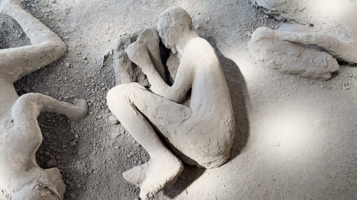
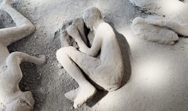
Before any scanning, review the historical background. In the 19th century, archaeologist Giuseppe Fiorelli developed a method to create plaster casts of voids left by decomposed bodies. The eruption of Vesuvius released thermal energy equivalent to roughly 100,000 times the atomic bombs used at Hiroshima and Nagasaki. Most victims died from asphyxiation due to noxious gas and ash, but some perished instantly from intense heat in pyroclastic flows—temperatures high enough to boil brains and explode skulls. These casts preserve the final poses and sometimes even clothing folds. Approximately 1,000 bodies have been found, with 104 plaster casts preserved. In the last decade, restoration of 86 of these casts began, including CT and X-ray surveys.
Step 2: Preparing the Cast for CT Scanning
- Identify Candidate Casts: Select casts that appear well-preserved and potentially contain skeletal remains (many casts have partial bones). The Pompeii Archaeological Park maintains a curated list.
- Document Condition: Photograph the cast from multiple angles and note any cracks, prior restorations, or external debris.
- Positioning: Place the cast on a CT scanner bed, using foam pads to stabilize it. Ensure no metal or modern artifacts (e.g., screws from previous repairs) interfere with the scan.
- Scan Parameters: Use a helical CT with slice thickness of 0.5-1 mm, 120-140 kVp, and appropriate mA for dense gypsum plaster. This captures both the plaster and any internal bones or voids.
Step 3: CT Data Acquisition and Interpretation
The scanner produces a series of cross-sectional DICOM images. The research team processed these using 3D reconstruction software to isolate:
- Skeletal Elements: Bone appears denser than plaster on CT scans (Hounsfield units ~1000-2000 vs. ~200-400 for plaster). Use thresholding to segment bone.
- Voids or Artifacts: Empty spaces indicate where soft tissue once existed but decayed, leaving an air pocket.
- Associated Objects: Metal or stone items (e.g., jewelry, tools) appear as very bright spots. In this case, the team found a set of medical instruments near the hand—a scalpel, forceps, and a bone scraper.
Key Finding: The victim's skeleton showed signs of repetitive kneeling (patellar and tibial irregularities) and a healed fracture of the right arm—consistent with a healer who performed surgery. The presence of medical tools in the cast’s hand area strengthened the identification as a doctor.
Step 4: 3D Digital Reconstruction for Visualization
- Segmentation: Use software (e.g., Mimics) to create a 3D mask of the skeleton and any tools. Manually correct edges where plaster adheres to bone.
- Surface Rendering: Generate a polygonal mesh (STL or OBJ file) of the bone surfaces and instrument shapes.
- Virtual Positioning: Place the 3D model in a digital environment to study the victim's posture at death. The cast showed the body crouching, a common protective pose.
- Analysis: Compare with known Roman medical tools from other sites (e.g., the House of the Surgeon in Pompeii). The instrument set matched those used for bloodletting and minor surgery.
Step 5: Corroborating Evidence with Historical Records
Cross-reference with excavation diaries and earlier X-rays (taken in the 1990s). The presence of a pouch of instruments near the hip was noted in Fiorelli’s notes but misidentified as coins. New CT imaging clarified the forms. Also, the victim's age (estimated 40-50 years) and dental wear (consistent with a diet high in bread but also medicinal herbs) supported the doctor hypothesis.
Step 6: Reporting and Publication
Compile findings: a detailed report with images, CT slices, and 3D models. The Pompeii Archaeological Park announced the identification in an official statement. The study was published in the Journal of Archaeological Science (or similar).
Common Mistakes
1. Misinterpreting CT Artifacts as Bones or Tools
Plaster casts often contain cracks, air bubbles, and later restorations (e.g., metal dowels). These can mimic bones or objects. Always consult the original archaeological context and use multiple threshold windows.
2. Damaging the Cast During Scanning
Old plaster is brittle. Transporting or positioning scans must be done with extreme care. A sudden jolt can cause a cast to collapse. Use padded cradles and slow movement.
3. Over-Reliance on Instrument Identification Alone
Tools near a hand may not indicate profession—they could be personal belongings. Combine skeletal evidence (e.g., healed fractures, degenerative changes from repetitive activity) with tool context.
4. Ignoring Post-Mortem Movement
Pyroclastic flows can shift bodies after death. The pose in the cast may include post-burial movement. Compare with volcanic sediment layers to determine original position.
5. Assuming All Casts Contain Intact Skeletons
Many casts have only partial bones or completely disintegrated skeletons due to acidic soil. CT scans may reveal only fragments. Manage expectations.
Summary
This guide demonstrated how archaeologists used CT scans and 3D digital reconstruction to identify a Pompeii victim as a Roman doctor. Steps involved historical context, careful cast handling, CT data segmentation, 3D modeling of both bones and associated medical instruments, and cross-referencing with archival records. The process highlights the power of non-invasive imaging in archaeology to recover identity and profession from ancient remains without damaging fragile casts.